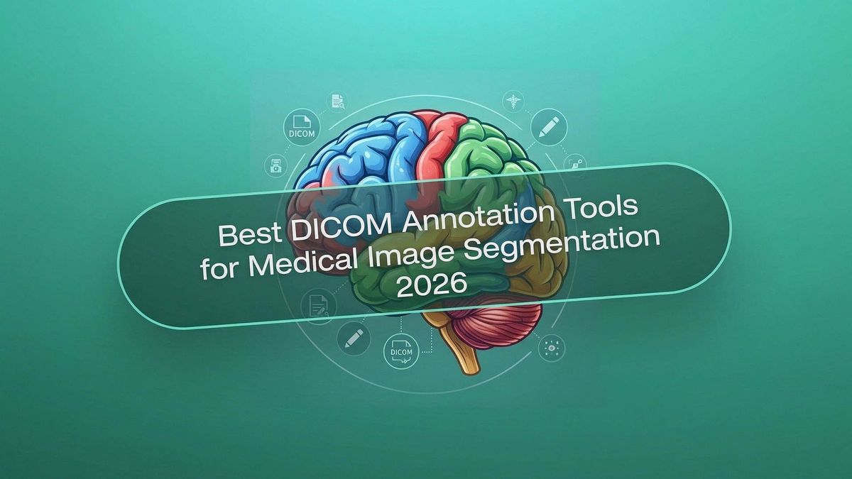
Best DICOM Tools for Medical Image Segmentation 2026
In 2026, the efficiency of diagnostics and surgical planning is inextricably linked to the quality of data preparation. At the heart of this ecosystem lies DICOM – a universal international standard that transforms an ordinary visual file into a complete medical document. Without understanding the specifics of this format, it is impossible to build any reliable artificial intelligence model capable of operating in clinical settings.
Annotation in medicine is the creation of standardized answers from which a neural network learns. A key stage in training medical AI is segmentation – the process of isolating specific anatomical objects or zones of interest on an image. While in classical computer vision, an error of a few pixels when labeling a car on a photo is negligible, in medicine, a margin of error within a millimeter during the segmentation of a vessel or a neoplasm can radically change treatment tactics. Therefore, DICOM tools in 2026 are complex systems that combine mathematical precision, medical expertise, and intelligent automation algorithms.
Quick Take
- The DICOM format preserves critical metadata, without which accurate medical diagnosis is impossible.
- Automatic contouring and smart interpolation reduce the radiologist's workload.
- The expert no longer draws masks manually but acts as the chief auditor of AI outputs.
- Professional platforms natively support HIPAA/GDPR and patient data anonymization.
Standards and Methods of Medical Labeling
Working with medical images is much more complex than ordinary photo processing. Every pixel here can affect human health; therefore, medical image labeling requires specialized software.
Technical Requirements for Professional Tools
Modern segmentation tools must be able to work with volumetric data because CT and MRI results consist of hundreds of slices that form a single 3D body. The program must have full DICOM support to preserve important patient information and machine settings. High precision for every isolated millimeter of tissue is also essential.
Important aspects include the safety and convenience of specialists' work:
- Healthcare compliance. Strict adherence to HIPAA and GDPR data protection rules to maintain patient confidentiality.
- Metadata handling. Automatic reading of information regarding slice thickness and body coordinates in space.
- Radiologist workflow. A convenient environment where several doctors can simultaneously verify each other's work and apply digital signatures.
- 3D volumes. The ability to view organs from any side rather than just on flat images.
Varieties of Segmentation in Medicine
For artificial intelligence to understand medical tasks, developers use different approaches to object isolation. Each method has its own purpose and assists doctors in specific situations. Choosing the correct type of labeling allows the model to better distinguish healthy tissues from pathologies.
Here are the main types of tasks solved by modern tools:
Thanks to these methods, artificial intelligence learns to see the body's structure as clearly as an experienced radiologist. This makes the diagnostic process faster, and treatment results much more reliable.
Technological Arsenal of Modern DICOM Tools
Modern tools for working with DICOM in 2026 are full-fledged ecosystems that minimize routine and maximize accuracy. When evaluating segmentation software, one should pay attention to six key functions that determine the speed and quality of medical data preparation.
- AI-assisted pre-labeling. Using pre-trained or custom models to automatically apply labels to the entire dataset before a specialist begins work.
- Auto-contours. A function for instantaneous isolation of organ boundaries or pathologies based on tissue density differences and anatomical atlases.
- Smart interpolation. Intelligent "filling" of gaps between slices. If you have placed labels on the first and tenth slices of a CT scan, the system automatically calculates the ideal contours for all layers between them, accounting for the object's anatomical shape.
- Model-in-the-loop annotation. A process where the AI model learns directly during labeling. Every correction made by a doctor instantly retrains the algorithm, making its subsequent suggestions more accurate.
- QA workflows. Multi-level verification systems where one doctor labels the data and another confirms its correctness. This creates the "double-check" necessary for medical certification.
- Version control for datasets. A "time machine" for data. It allows seeing who made changes and when, comparing different versions of annotations, and returning to previous versions if a new model shows worse results.
These functions transform an annotation tool into an intelligent assistant, allowing radiologists to focus on complex clinical cases while delegating technical work to algorithms.
Overview of Top DICOM Tools 2026
In 2026, the DICOM tool market is clearly divided into two camps: powerful desktop platforms for deep scientific research and cloud-based enterprise solutions for rapid scaling of AI projects. Let's compare the leaders defining industry standards.
3D Slicer
The most versatile free platform for medical visualizations. Thanks to thousands of plugins, it remains the "gold standard" in the academic environment.
- Strengths: enormous library of extensions, VR/AR support, and full compatibility with NVIDIA's MONAI framework.
- Use Cases: complex scientific research, prototyping new treatment methods, and surgical planning.
- Status: research.
RedBrick AI
A modern SaaS platform created specifically for preparing medical data at scale. It focuses on making the labeling process as fast as possible for teams.
- Strengths: cloud architecture, built-in tools for automatic 3D segmentation, and convenient management of medical teams.
- Use Cases: training commercial AI models where rapid processing of thousands of CT/MRI scans is required.
- Status: enterprise.
ITK-SNAP
A highly specialized tool that does one thing but does it flawlessly – segmenting 3D structures.
- Strengths: incredibly stable "active contours" algorithm and interface simplicity.
- Use Cases: fast manual or semi-automatic segmentation of specific organs or tumors on brain or heart MRIs.
- Status: research.
Encord
A platform that grew out of classical computer vision but became a leader in medical video and dynamic DICOM data.
- Strengths: best support for 4D and a powerful SDK for developers.
- Use Cases: projects where it is important to track object changes over time.
- Status: enterprise.
OHIF Viewer
A web framework that can be embedded into any browser.
- Strengths: operates in the browser without delays, easily integrates with hospital systems.
- Use Cases: quick viewing of images in a clinic and light annotation "on the fly".
- Status: hybrid (used in both development and clinics).
Keylabs
A professional annotation platform combining powerful segmentation tools with flexible project management. It is ideal for creating high-precision datasets where the quality of every pixel is decisive.
- Strengths: native DICOM support, smart interpolation between slices, and a built-in quality control system allowing professional radiologists to be involved in validation.
- Use Cases: creating "gold standard" data for diagnostic AI systems in narrow domains.
- Status: enterprise.
Open-source & Enterprise
The choice of tool depends on the stage of your project. If you are looking for a new mathematical model, you need flexibility. If you are building a product for thousands of hospitals, you need standards and security.
Main Differences by Categories
1. Scope of Application
- Open-source (3D Slicer, ITK-SNAP). Ideal environment for research labs and universities. This is where new algorithms are born. Scientists can modify the code, add their own formulas, and test hypotheses without license restrictions.
- Enterprise (Keylabs, RedBrick AI, Encord). Targeted at Startups and large corporations. The speed of preparing millions of frames for model training is more important than the ability to change code. This is an industrial conveyor for AI.
2. Regulation and Security
- Hospitals and Regulated Environments. Hospitals and government institutions usually choose an enterprise due to healthcare compliance. Paid platforms already have certifications and provide encryption and logging of every action. In open-source, the user is responsible for data security, which carries significant legal risks.
Comparison of Trade-offs
The Future of Medical Segmentation
The technology stack of 2026 has finally formed three fundamental trends that define how medical AI products will be developed in the next decade.
The Era of Semi-automation
Thanks to foundation models, segmentation has become semi-automatic.
- Prompt-based segmentation. The doctor only points to an object or describes it with text, and the system instantly builds a 3D mask.
- Interactive refinement. The AI proposes a version, and the human only makes slight corrections, which reduces working time.
Changing Role of the Doctor
The role of the medical expert is shifting toward validation. Today, the doctor's main task in an AI project is expert supervision and correcting complex cases where automation might fail.
- Human-in-the-loop. The human becomes the final quality filter, ensuring patient safety.
- Expert supervision. Annotation platforms build their interfaces around the verification and approval process.
Data Quality Over Volume
The industry has realized that 1,000 perfectly segmented scans with verified diagnoses yield better results than 100,000 "dirty" annotations.
- Data-centric AI. The focus has shifted to finding and fixing errors in datasets.
- Gold standard datasets. Creating reference samples verified by multiple experts has become the primary value for model certification by regulators.
FAQ
How do segmentation tools help in the 3D printing of organs?
Segmentation transforms DICOM data into 3D models, which serve as the basis for printing personalized implants or surgical templates. The precision of the tool at this stage determines how perfectly the implant will fit the patient during surgery.
Why is it impossible to simply convert DICOM to JPG for AI training?
During conversion, color depth and metadata about the physical dimensions of voxels are lost, which makes it impossible to calculate the volume or density of tissue. Without these parameters, artificial intelligence will be unable to distinguish, for example, a cyst from a tumor.
How are disputes resolved if two doctors have segmented the same tumor differently?
In professional QA processes, a consensus method is used, or the involvement of a third "judge" with higher qualifications. Also, there are algorithms that automatically calculate the most probable boundary of an object based on several opinions.
Are there special requirements for the monitors on which annotators work?
Yes, for medical labeling, calibrated monitors are required that comply with the DICOM standard for the correct display of gray shades. This guarantees that the annotator sees the same tissue details that a diagnostic doctor would see in a clinic.
What is the role of synthetic data in medical segmentation?
Synthetic data helps to "complete" samples for rare diseases where real clinical cases are too few for training a neural network. However, such data always undergo strict validation by doctors so that the AI does not begin to invent non-existent anatomical anomalies.
How is data security ensured when using cloud platforms?
Data are encrypted, and all identification information of the patient is automatically removed from the file metadata even before it gets into the cloud. This allows teams to work remotely without violating laws on medical secrecy.
Are programming skills needed to work with professional segmentation tools?
For most enterprise solutions, programming is not needed since they have an intuitive interface. However, for open-source tools, knowledge of Python will help automate specific tasks and install complex plugins.

Comments ()